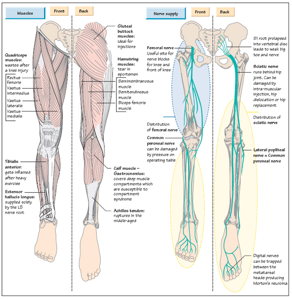
Anatomy Of Upper Thigh And Hip - Thigh & Hip / Quadriceps, a group of four.. It is part of the lower limb. Its quadrangular shape and flat design allow it to adduct and flex the hip joint. Knowing the anatomy of your hip can help you understand the source of any hip pain. Several muscles cross the front of the hip and create hip flexion, pulling the thigh and trunk toward both muscles cross the floor of the pelvis, emerge at the outer edges of the pubic bones, and finally insert on the inner upper femur (thighbone). The adductor muscle on the inner thigh;
Foundational anatomy provides medical students with the necessary background in anatomy for success in clerkships. The iliopsoas muscle, which extends from the lower back to upper femur; Jew anatomy atlases, the anatomy atlases logo, and a digital library of anatomy information are all trademarks of michael p. Mri of upper leg (femur). This webpage presents the anatomical structures found on hip mri.
Pin on Body Moves - To the Point from i.pinimg.com The upper part of the thigh bone consists of the femoral head, femoral. Bones of the lower limb. Tibial part of the sciatic nerve action: The femur or thigh bone is one of the longest bones in the human body. He also serves the communities of charleston, sc and augusta, ga. This mri hip joint axial cross sectional anatomy tool is absolutely free to use. The single bone in the thigh region is called the origin: Pelvis, perineum, hip, and upper thigh.
In vertebrate anatomy, hip (or coxa in medical terminology) refers to either an anatomical region or a joint.
It functions to adduct the thigh and to flex. It is part of the lower limb. The hip muscles are going to be slip into hip muscles and gluteal muscles. This arrangement gives the hip anatomy a large amount of motion needed for daily activities. The thigh is the area between the hip and the knee joint. The iliopsoas muscle, which extends from the lower back to upper femur; While the thigh muscles will be slip into the anterior, medial and posterior groups. May 13, 2019 edited by dr. Tibial part of the sciatic nerve action: Quadriceps, a group of four. Medial condyle of tibia nerve supply: Unlike the shoulder girdle, the pelvic girdle is firmly integrated into the axial skeleton: Hip surgeon dr guillaume dumont offers hip pain treatments in columbia, sc.
Hip movements include flexion, extension, abduction, adduction, circumduction, and hip rotation. Foundational anatomy provides medical students with the necessary background in anatomy for success in clerkships. Hip flexor deep in pelvis a composite o… used to extend the hip when climbing st… The uppermost of the medial thigh muscles is the pectineus muscle. This vein, as well as the deep veins.
Anatomy of the Leg | Musculoskeletal Key from musculoskeletalkey.com It is part of the lower limb. Each pelvic girdle consists of a hip bone (coxal bone, innominate bone), which articulates with the head of a femur. This deep muscle begins in the low back and pelvis and connects on the inside edge of the upper femur. A, anterior and posterior views show the hip joint ligaments. While the thigh muscles will be slip into the anterior, medial and posterior groups. Muscles of the hips and thighs. The thigh is the area between the hip and the knee joint. Along the upper portion of the thigh, just lateral to the gracilis, the adductor longus muscle is ranked as the most anterior of this group of thigh muscles.
Hip anatomy, function and common problems.
This mri hip joint axial cross sectional anatomy tool is absolutely free to use. While the thigh muscles will be slip into the anterior, medial and posterior groups. In vertebrate anatomy, hip (or coxa in medical terminology) refers to either an anatomical region or a joint. The muscles also require a lot of blood flow, which provides oxygen and nourishment, especially when you're physically active. The hip joint is a ball and socket joint that is the point of articulation between the head of the femur and the acetabulum of the pelvis. Pelvis, perineum, hip, and upper thigh. Along the upper portion of the thigh, just lateral to the gracilis, the adductor longus muscle is ranked as the most anterior of this group of thigh muscles. Like the forearm, the upper leg, or thigh, has a dense arrangement of many muscles. for detailed anatomy of pelvic bones, read anatomy of hip bone. Medial condyle of tibia nerve supply: All of the anatomical parts of the hip work together to enable various movements. The iliopsoas muscle, which extends from the lower back to upper femur; Hip surgeon dr guillaume dumont offers hip pain treatments in columbia, sc.
Anatomy ▶ lower limb ▶ bones and cartilages ▶ hip joint. The upper part of the thigh bone consists of the femoral head, femoral neck, and greater and lesser trochanters. Groin, inguinal region and the anterior and posterior regions of the hip and thigh. Pelvis, perineum, hip, and upper thigh. Superficial fascia.—the superficial fascia forms a continuous layer over the whole of the thigh;
Muscles of the Thigh Part 2 - Medial Compartment - Anatomy ... from i.ytimg.com A, anterior and posterior views show the hip joint ligaments. The hip muscles are going to be slip into hip muscles and gluteal muscles. 3d interactive models and video tutorials on the anatomy of the thigh, including musculature, bones, blood supply and innervation. Use the mouse scroll wheel to move the images up and down alternatively use the tiny arrows (>>) on both side of the image to move the images. Anatomy ▶ lower limb ▶ bones and cartilages ▶ hip joint. This webpage presents the anatomical structures found on thigh mri. Tibial part of the sciatic nerve action: He also serves the communities of charleston, sc and augusta, ga.
Jew anatomy atlases, the anatomy atlases logo, and a digital library of anatomy information are all trademarks of michael p.
for detailed anatomy of pelvic bones, read anatomy of hip bone. 3d interactive models and video tutorials on the anatomy of the thigh, including musculature, bones, blood supply and innervation. The joints and muscles of the hips and thighs need nervous input so they can do what your brain wants them to do. Anatomy hip, thigh and leg muscles. Quadriceps, a group of four. Pelvis, perineum, hip, and upper thigh. The following nerves serve the gluteal and. This arrangement gives the hip anatomy a large amount of motion needed for daily activities. This vein, as well as the deep veins. Groin, inguinal region and the anterior and posterior regions of the hip and thigh. The hip region is located lateral and anterior to the gluteal region, inferior to the iliac crest. Hip and knee pain and hip and shoulder pain are. Anatomy of the human body.


0 Komentar